Anatomy Of Musckes Sndctendons | Cardiac muscle contracts the heart to pump blood. There are around 650 skeletal muscles within the typical human body. Each of these muscles is a discrete organ constructed of skeletal muscle tissue, blood vessels, tendons, and nerves. See anatomy pictures of the 27 bones in the hand and wrist, how they are connected with tendons and muscles and the nerves that run through the skeletal structure. Rectus capitis, longus capitis, longus colli.
As the skeletal muscles pull on bones to cause movements, they also stabilize the joints of the skeleton; Human muscle system, the muscles of the human body that work the skeletal system, that are under voluntary control, and that are concerned with the following sections provide a basic framework for the understanding of gross human muscular anatomy, with descriptions of the large muscle groups. This is a table of skeletal muscles of the human anatomy. Cardiac muscle contracts the heart to pump blood. These muscles originate from the surface of the skull and insert onto the mandible.¹.
The tendons of these muscles pass through a small corridor in the wrist known as the carpal tunnel. How to study muscle anatomy. Muscular contraction is necessary for voluntary and involuntary movement of limbs, stabilization of joints, maintaining luminal diameter (in the case of arteries, bowel, etc), and to produce heat. Inflammation of this region caused by repetitive stress or trauma may lead to pain and numbness known as carpal tunnel syndrome. Attached to the bones of the skeletal system are about 700 named muscles that make up roughly half. Each of these muscles is a discrete organ constructed of skeletal muscle tissue, blood vessels, tendons, and nerves. However, if you take a little time to learn a few root words, those latin names can give you valuable insights into things like the muscle's size and shape. It elevates and protrudes the mandible. Digastric, mylohyoid, geniohyoid, stylohyoid infrahyoid muscles: Long flexor tendons extend from the forearm muscles through the wrist and attach to the small bones of the fingers and thumb. Anterior muscles of the neck. Related online courses on physioplus. • muscle tissues develop from embryonic cells.
In this section, learn more about the anatomy of the muscles of the neck. The anterior and middle scalenes originate from the transverse processes of certain cervical vertebrae and attach to the first rib. An interactive tutorial teaching the position, actions, innervation and attachments of the rectus femoris muscle with the aid of anatomical illustrations. See anatomy pictures of the 27 bones in the hand and wrist, how they are connected with tendons and muscles and the nerves that run through the skeletal structure. The muscular system is responsible for the movement of the human body.

This is a table of skeletal muscles of the human anatomy. Skeletal muscle moves bones and other structures. There are four muscles that comprise the muscles of mastication. Long flexor tendons extend from the forearm muscles through the wrist and attach to the small bones of the fingers and thumb. Sternohyoid, sternothyroid, thyrohyoid, omohyoid anterior vertebral muscles: Leg anatomy muscle anatomy anatomy study anatomy reference psoas release anatomy models muscular system medical anatomy human the muscles of the shoulder and back chart shows how the many layers of muscle in the shoulder and back are intertwined with the other. As the skeletal muscles pull on bones to cause movements, they also stabilize the joints of the skeleton; Inflammation of this region caused by repetitive stress or trauma may lead to pain and numbness known as carpal tunnel syndrome. The muscular system is responsible for the movement of the human body. Understanding the structure of a muscle fiber. In the muscular system, muscle tissue is categorized into three distinct types: Microscopic anatomy of skeletal muscle. • the muscular system develops from intra embryonic mesoderm.
Sternohyoid, sternothyroid, thyrohyoid, omohyoid anterior vertebral muscles: Muscle movements, types, and names. It elevates and protrudes the mandible. Muscles are tissues that contract to help parts of the body move. The muscles of the torso, examined in the previous chapter, include a few that attach directly into the upper arm and help move the humerus at the shoulder joint.
Movement of the mandible at the temporomandibular joint). The muscular system is responsible for the movement of the human body. Anatomy of the muscular system. Cardiac muscle contracts the heart to pump blood. Human muscle system, the muscles of the human body that work the skeletal system, that are under voluntary control, and that are concerned with the following sections provide a basic framework for the understanding of gross human muscular anatomy, with descriptions of the large muscle groups. Leg anatomy muscle anatomy anatomy study anatomy reference psoas release anatomy models muscular system medical anatomy human the muscles of the shoulder and back chart shows how the many layers of muscle in the shoulder and back are intertwined with the other. This is a table of skeletal muscles of the human anatomy. The three scalene muscles are found forming the floor of the posterior triangle. Along with lateral pterygoid muscle it produces side to side movement of mandible. Topographically, the muscles in this group are classed along with the lateral torso wall and upper extremity, which is due to their location as well as their genetic development based on their embryological origin. Understanding the structure of a muscle fiber. Long flexor tendons extend from the forearm muscles through the wrist and attach to the small bones of the fingers and thumb. Muscle mass accounts for a large majority of the carcass weight of domestic animals.
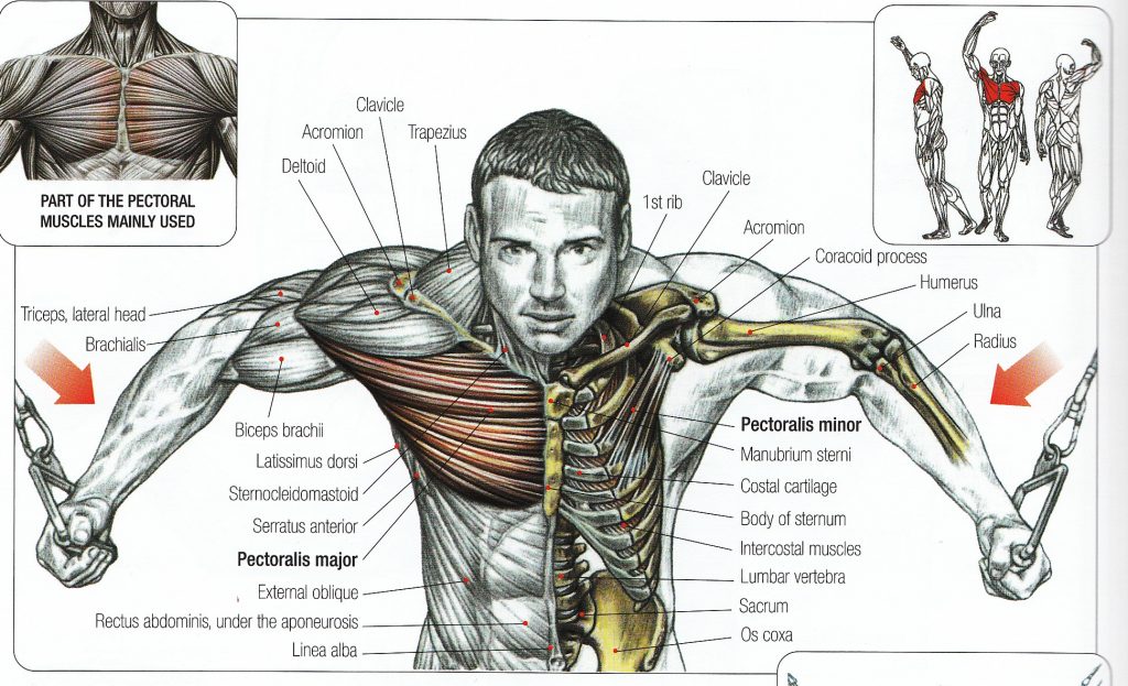
Anatomy Of Musckes Sndctendons: Anatomy of the short head of the biceps brachii muscle.